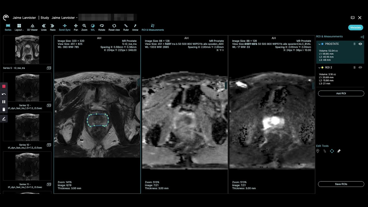
Are you looking for how to read prostate MRI images? Then this article is just right for you! This article gives a brief introduction on how to read prostate MRI images. Specifically, we’ll discuss the 2 common methods of imaging prostate cancer, and about the applications of various methods of prostate imaging, including MRIs, PIRS, and ultrasound guided tomography (GDT). Additionally, we’ll talk about related diagnostic procedures that are used together with these prostate cancer imaging modalities.
One of the most common techniques of prostate imaging is the digital rectal exam (DRE) and prostate specific antigen (PSA) detection. The goal of the DRE is to detect an abnormal mass or tumor in the transrectal canal or the seminal vena cava using magnetic resonance imaging technology. The main tool employed in the DRE is the digital rectal exam probe, which has been well-known for its high sensitivity and high success rate in prostate cancer detection. Consequently, many prostate urologists need to use DRE to assess the state of their patients’ prostate cancers.
Another prostate imaging modality is the prostate-specific antigen scan or PSA. The PSA is used to measure the levels of PSA in a patient’s blood. This method has a number of health benefits for cancer patients, including early detection of cancer. Although the PSA does not directly diagnose prostate cancer, it can help to determine if there are any changes in the prostate such as a tumor or growth. Typically, the PSA scan is done in a doctor’s office and is more sensitive than the DRE. In addition, it has a faster speed and smaller images.
The last non-invasive method in learning how to read prostate MRI is called the transrectal ultrasound or TRUS. This technology uses sound waves to send ultra-thin bubbles through the urinary tract to visualize the interior of the bladder. A special instrument called micropores is used to send the sound waves through the urethra. Because the bubbles reflect light, they form images that are used to create a contrast in color. In order to create an accurate image, several smaller bubbles must be sent at once.
There are some key benefits to this prostate cancer imaging modality. The first is that it can provide a quick look inside the bladder and prostate to identify abnormal cells without requiring an overnight stay in the hospital. This minimally invasive procedure also offers a high level of accuracy, which makes it a popular choice for prostate cancer detection. TRUS does not destroy abnormal prostate cells, but it does observe them. In fact, some small prostate cancers can be seen using this technique as well.
As mentioned, one of the benefits of learning how to read prostate cancer from MRI is that it allows men with prostate problems to find out whether or not they have any abnormalities. It can also help them discover potential imbalances. If you suffer from any pain, you might find that you can tell from looking at your lower back and pelvis. This can help you make a more informed decision about the best course of treatment.
Another advantage to this type of prostate cancer imaging is that the procedure is painless. You can find yourself more comfortable during the procedure. Your doctor may suggest that you go home with a warm bath and a pillow or two to take your mind off the discomfort of the scan. After a few hours, you will most likely find yourself feeling much better.
Learning how to read prostate cancer MRI scans can help you decide if there is something wrong. You don’t want to wait for more tests before seeing your physician. Don’t be afraid to ask questions. You never know what you are suffering from. You might find that learning how to read prostate cancer MRI scans is exactly what you need to move forward with your health.
Source:
